Common Retinal Disorders
LEARN MORE
Learn About the most common disorders we treat below
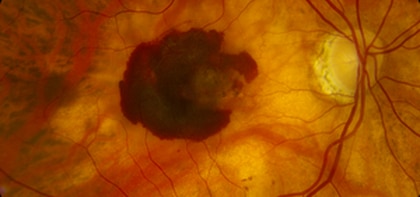
Age-Related Macular Degeneration
(AMD) is a deterioration of the retina and
choroid that leads to a substantial loss in visual acuity (sharpness of vision). AMD is the leading cause of significant visual acuity loss in people over age 50 in developed countries.

Diabetic Retinopathy
Diabetic retinopathy is a complication of diabetes that causes damage to the blood vessels of the retina— the light-sensitive tissue that lines the back part of the eye, allowing you to see fine detail.
Diabetic retinopathy is the most common cause of irreversible blindness in working-age Americans. As many people with type 1 diabetes suffer blindness as those with the more common type 2 disease. Diabetic retinopathy occurs in more than half of the people who develop diabetes.
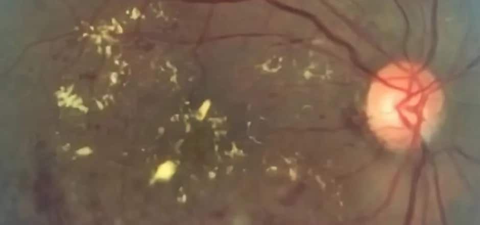
Macular Edema
The macula is the part of the retina that helps us see fine detail, faraway objects, and color. It’s packed with more photoreceptors (light-sensitive cells) than any TV or monitor. The small, central area of the retina is worth the most—the bullseye of sight. Macular edema, degeneration, hole, pucker, drusen (small yellowish deposits), scar, fibrosis, hemorrhage, and vitreomacular traction are common conditions that involve the macula. When macular disease is present, distorted vision (metamorphopsia), blank spots (scotoma), and blurred vision are common symptoms.
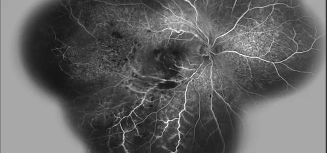
Branch Retinal Vein Occlusion
Retinal vein occlusions occur when there is a blockage of veins carrying blood with needed oxygen and nutrients away from the nerve cells in the retina. A blockage in the retina’s main vein is referred to as a central retinal vein occlusion (CRVO), while a blockage in a smaller vein is called a branch retinal vein occlusion (BRVO).

Central Retinal Vein Occlusion
Central retinal vein occlusion, also known as CRVO, is a condition in which the main vein that drains blood from the retina closes off partially or completely. This can cause blurred vision and other problems with the eye.
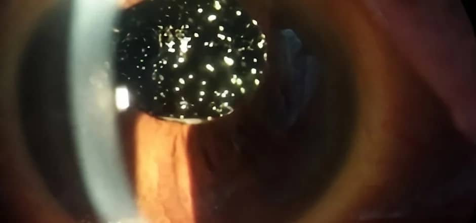
Vitrectromy for Floaters
As we get older, some of us may see
"floaters" in our field of vision. In rare cases when many of these floating specks interfere with vision while driving or reading, one treatment option is vitrectomy—surgical removal of the eye’s vitreous gel.
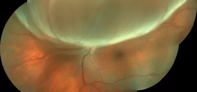
Retinal Detachment
The retina lines the back wall of the eye, and is responsible for absorbing the light that enters the eye and converting it into an electrical signal that is sent to the brain via the optic nerve, allowing you to see. Many conditions can lead to a retinal detachment, in which the retina separates from the back wall of the eye, like wallpaper peeling off a wall.
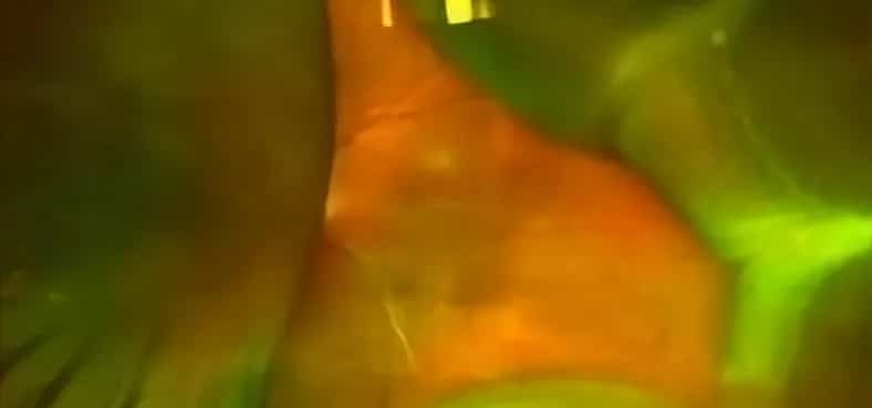
Choroidal Detachment
The choroid (pronounced “CORE-oyd”) is a spongy layer of blood vessels that lines the back wall of the eye between the retina and the sclera (or the white part of the eye). It plays an important role in delivering oxygen and nutrients to the outer half of the retina. The choroid is normally directly next to the sclera, but can be displaced by fluid or blood, leading to a choroidal detachment (separation).

Central Serous Chorioretinopathy
Central serous chorioretinopathy, commonly referred to as CSC, is a condition in which fluid accumulates under the retina, causing a serous (fluid-filled) detachment and vision loss.
CSC most often occurs in young and middle-aged adults. For unknown reasons, men develop this condition more commonly than women. Vision loss is usually temporary but sometimes can become chronic or recur.